Wound Assessment
Wound assessment is the key to successful wound management. A detailed and accurate description of the etiology of the wound, the wound itself, and the plan of care is key to the successful management of wounds. The acronym T.I.M.E. has been successfully used to assist in writing wound descriptions.
T is for Tissue |
wound bed color helps to determine if there is unhealthy tissue present |
I is for Infection |
is there bacteria in the wound that could impeded healing by delaying the inflammatory phase. |
M is for Moisture |
a damp environment is best for healing when the wound be is dry the growth factors are unable to activate granulation and angiogenesis and if it is to moist the growth factors are weakened. |
E is for Edge |
if the wound is to moist the edge and surrounding tissue can become macerated. Tissue damage can occur inhibiting wound healing. (Brawn, 2020) |
Health History
A persons' health history is a crucial component to wound management. Comorbidities, such as diabetes, vascular disease, and nutritional status, are important factors in wound healing.
For example, diabetes is associated with venous and arterial insufficiency, which results in reduced blood flow needed for wound healing. Nutrition and hydration can also impact wound healing. Reviewing medications, including over-the-counter medications, and herbal and dietary supplements is also essential.
Labs appropriate for wound care (Hess, 2015)
Albumin is a protein that acts as a building block for cells and tissues. Albumin is the primary screening tool for protein status and is a gross indicator of nutritional status and fluid balance.
Prealbumin, or transthyretin, is another type of protein produced by the liver. ... It has a half-life of 2 to 3 days, making it a better indicator of acute nutritional status changes than albumin.
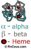
 Hemoglobin A1c. is composed of hemoglobin Alpha with a glucose molecule, which is attached through a process called glycosylation. HGA1c is an indicator of long-term glucose control. It is frequently used in the management of diabetes because of its link to elevated glucose levels.
Glucose is formed from dietary carbohydrates and is stored in the liver and muscles as glycogen. Glucose is frequently elevated in persons with diabetes, renal failure, and persons taking steroid medication. Elevated glucose impairs wound healing.
A complete blood count (CBC) measures the number of red blood cells, white blood cells, the total amount of hemoglobin in the blood, the fraction of the blood composed of red blood cells (hematocrit), and the mean corpuscular volume. It is essential to review these blood components because they map directly to the wound-healing process.
Instant Feedback:
Albumin is a protein that acts as a building block for cells and tissues
Location and type of wound
Where is the wound located, and what is the etiology of the wound? You must be as clear and detailed as possible. Anatomical positions and landmarks can be helpful to accurately describe the location of the wound. Visual aids and pictures can be beneficial. If the wound is over a bony prominence, then it is usually a pressure wound. Wounds on the lower extremities are typically venous, arterial, and or diabetic ulcers.
Description and classification
Wounds are generally identified by the degree of tissue damage. Tissue layers involved partial or full-thickness. The color of the tissue in the wound is also essential. Is it yellow sloughing or pink? Is there eschar present. One of the most common types of wounds present is the pressure injury.
Pressure injuries involve the skin and underlying tissue. The National Pressure Injury Advisory Panel (NPIAP; formerly the National Pressure Ulcer Advisory Panel [NPUAP]) identify a pressure injury to be localized damage to the skin and underlying soft tissue, usually over a bony prominence. It occurs as a result of intense or prolonged pressure or pressure in combination with shear (Kirman, 2020).
NPUAP Stages of a Pressure Ulcer |
Stage 1 pressure injury - |
The skin is intact. The erythema in a localized area and non-blanchable |
In people with dark skin tones, stage one may be difficult to identify. The skin may appear dusky or grayish. |
Stage 2 pressure injury - |
There is a Partial-thickness skin loss with exposed dermis. This may be a shallow open area with a red-pink wound bed |
|
Stage 3 pressure injury - |
There is full-thickness skin loss. Subcutaneous fat I may be visible. |
May include undermining or tunneling |
Stage 4 pressure injury - |
Full-thickness skin and tissue loss, bone exposed as well as muscle and tendons |
|
Unstageable pressure injury - Obscured full-thickness skin and tissue loss |
There is full-thickness loss, slough, and eschar cover the base of the wound |
|
Deep pressure injury |
The skin color is deep red, maroon or purple discoloration It is Persistent and non-blanchable |
Initially, the area may be painful, boggy, or tender, or with blisters, fluid-filled with blood |
Measurements
Wound measurements are the key to evaluating wound healing. All members of the wound care team should follow the same procedure for measuring a wound. Wound measurements should be documented in centimeters and done consistently, to increase accuracy and reliability. Wounds are three -dimensional with (length) X (width) X (depth) measurements. Photographs of the wound should be done regularly to identify healing and delays.
Measuring undermining and tunneling
It is crucial to identify undermining and tunneling. Undermining occurs when the tissue under the edges of the wound becomes eroded, causing a lip or pocket to the wound. The area is measured by gently placing a probe or a moistened cotton tip applicator under the edge of the wound. The probe or applicator is marked where it meets the wound edge.
Tunneling occurs when a narrow tract or pathway of tissue damage is formed deeper into the subcutaneous tissue. Tunneling can happen in the presence of bacteria and improper packing of wounds, such as dressings that dehydrate the wound and create constant pressure.
The location of the tract within the wound must be documented. The clock model is helpful in the description. Superimpose the face of a clock over the wound. The patient's head is 12 o'clock and the feet at 6 o'clock. For example, the hip wound is 8cm X 5cm X 2cm with a tract measuring 6 cm at 3 o'clock laterally.
Drainage and Odor
Drainage or exudate is evaluated based on the amount color, odor, and consistency.
Amount - scant, minimal, moderate, or heavy?
Color - opaque, clear serosanguinous, bloody, red, tan, brown, yellow, and green?
Odor - foul-smelling odor or sweet scent? Does cleansing eliminate or decrease the odor?
Consistency is also important, is the exudate thin, watery, creamy, or thick?
For example, a healing wound will have a red wound bed with clear serosanguineous exudate.
Wound edges are part of the wound description. They are usually described as rounded firm, elevated, flat, or edematous. The skin surrounding the wound is equally essential. Is it warm to touch red, hot pale, or cold? If it is erythematous, how far does the erythema extend? Note if it is less than 5 cm or greater.
A photo journal of the wound is extremely helpful in monitoring progress and identifying problems, photograph the wound head-on, not at an angle. The first photography should be of the entire area of the body where the wound is present. Try to get close to the wound no further than 6-10 inches away transfer the photo to the EMR (WOCN, Board of Directors Task Force, 2020).
Instant Feedback:
Wounds are three-dimensional with length X width X depth measurements.
References
Brawn, K. (2020) Guidelines for the Assessment & Management of Wounds (rev. 12/2022) retrieved from https://www.nhft.nhs.uk/download.cfm?ver=51721
Hess, C. T. (2015). Auditing Wound Care Documentation. Advances in skin & wound care 28(5), 240. DOI:10.1097/01.ASW.0000464707.80878.68
Hess, C. T. (2015). Clinical Order Sets: Defining Lab Tests for Wound Care. Advances in Skin & Wound Care 28(3), 144. DOI:10.1097/01.ASW.0000461295.42250.ec
Kirman, C. (2020). Pressure Injuries (Pressure Ulcers) and Wound Care Retrieved from https://emedicine.medscape.com/article/190115-overview
NPUAP pressure injury stages. (2016). National Pressure Injury Advisory Panel. Available at https://cdn.ymaws.com/npiap.com/resource/resmgr/npuap_pressure_injury_stages.pdf.
Edsberg, L. E., Black, J. M., Goldberg, M., McNichol, L., Moore, L., & Sieggreen, M. (2016). Revised National Pressure Ulcer Advisory Panel Pressure Injury Staging System. J Wound Ostomy Continence Nurs, 43(6), 585-597. DOI:10.1097/won.0000000000000281
WOCN Board of Directors Task Force 9 (2020). Recommendations for wound assessment and photo-documentation in isolation. Journal of Wound Ostomy & Continence Nursing 47(4) 319-320.
©RnCeus.com
 Hemoglobin A1c. is composed of hemoglobin Alpha with a glucose molecule, which is attached through a process called glycosylation. HGA1c is an indicator of long-term glucose control. It is frequently used in the management of diabetes because of its link to elevated glucose levels.