Etiology of VSD
Congenital heart disease (CHD) occurs when something disrupts the normal development of the heart. Genetic and or environmental risks can influence the incidence of CHD. Most cases of CHD have their beginnings in weeks 4-8 of pregnancy (NHS. n.d.) when endothelial tube that develops and acquires the shape and structures of the chambered heart (RnCeus n.d.).
 Congenital septal defects, whether atrial or ventricular, result from abnormal growth and incomplete development of the atrial septum that divides the upper chamber into right and left atrial chamber or the interventricular septum dividing the lower chamber into right and left ventricles.
Genetic risk VSD factors include:
- VSD occurs more frequently in males than females
- Familial history of congenital heart disease
- Down syndrome (trisome syndromes)
- VSD may occur in association with a variety of other rare genetic disorders including Holt-Oram Syndrome, FG Syndrome, Genitopalatocardiac Syndrome, Fryns Syndrome, certain forms of Dandy-Walker Syndrome, Cardiomyopathy-Hypogonadism-Collagenoma Syndrome, Familial Idiopathic Cardiomyopathy, Simpson Dysmorphia Syndrome, and Velocardiofacial Syndrome (NORD (n.d.)).
Environmental factors include:
- Maternal diabetes or phenylketonuria
- Tabib, A., Shirzad, N., Sheikhbahaei, S., Mohammadi, S., Qorbani, M., et al. (2013). Cardiac malformations in fetuses of gestational and pre gestational diabetic mothers. Iranian journal of pediatrics, 23(6), 664–668.
- Levy HL, Guldberg P, Güttler F, Hanley WB, Matalon R,, et al. (2001). Congenital heart disease in maternal phenylketonuria: report from the Maternal PKU Collaborative Study. Pediatr Res. 49(5):636-42. doi: 10.1203/00006450-200105000-00005. PMID: 11328945.
- Exposure to disease or teratogens within in the first 8 weeks of gestation.
- Teratogens and congenital heart disease - Tara A. Lynch ... (n.d.). Retrieved April 21, 2022, from https://journals.sagepub.com/doi/full/10.1177/8756479315598524
- Maternal smoking and alcohol use effects on VSD prevalence is inconlusive
- Sands, A., Casey, F., Craig, B. et al. (1998). Ventricular Septal Defect; A Frequent Finding In Clinically Normal New-borns. Pediatr Res 44, 422 . https://doi.org/10.1203/00006450-199809000-00050
- Yang, J., Qiu, H., Qu, P., Zhang, R., Zeng, L., & Yan, H. (2015). Prenatal Alcohol Exposure and Congenital Heart Defects: A Meta-Analysis. PloS one, 10(6), e0130681. https://doi.org/10.1371/journal.pone.0130681
- Taylor, K., Elhakeem, A., Thorbjørnsrud Nader, J. L., Yang, T. C., Isaevska, et al. (2021). Effect of maternal prepregnancy/early‐pregnancy body mass index and pregnancy smoking and alcohol on congenital heart diseases: A parental negative control study. Journal of the American Heart Association, 10(11). https://doi.org/10.1161/jaha.120.020051
- Parent/family education should include the complexity of cardiac development and medical sciences's limited ability to ascertain causality.
Developmental errors
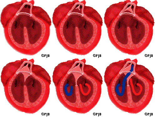
A VSD may result if cells from ventricle, endocardial cushions or truncus arteriosus fail to grow, organize or fuse to the appropriate structures. For example an intact membranous septum requires growth and fusion of tissues derived from the endocardial cushions, the conotruncal ridges and muscular septum. Errors in the growth rate, orientation or fusion of any of these tissues can result in VSD.
 Endocardial cushions
The first division is accomplished by the endocardial cushions. The endocardial cushions begin the separation of the heart into right and left, upper and lower chambers. These chambers will become the atria and ventricles. While the endocardial cushions continue to develop, the atrial and ventricular septa begin to form.
Muscular Septum
The ventricular septum is composed of thick, trabeculated muscular tissue derived from the walls of the growing ventricles. When this muscular septum is complete, a large opening (the interventricular foramen) still remains between the two ventricles. The interventricular foramen is usually completely closed by week seven. Closure is accomplished by growth of membraneous tissue derived from the endocardial cushions, the interventricular septum and from the conus ridges growing within the truncus.
 Membranous septum
The membranous septum derived from tissue growing within the truncus. The truncus begins to divide about 29 days after conception. The division starts with the growth of two ridges that take a spiral path toward the ventricles. The spirals eventually joining with tissue from the interventricular septum and closing the interventricular foramen. This tissue completes the separation of venous from arterial blood flow.
References
Lin KY, D'Alessandro LC, Goldmuntz E. (2013). Genetic testing in congenital heart disease: ethical considerations. World J Pediatr Congenit Heart Surg. 4:53–7. doi: 10.1177/2150135112459523
Ko J. M. (2015). Genetic Syndromes associated with Congenital Heart Disease. Korean circulation journal, 45(5), 357–361. https://doi.org/10.4070/kcj.2015.45.5.357
Kovalenko, A. A., Anda, E. E., Odland, J. Ø., Nieboer, E., Brenn, T., & Krettek, A. (2018, June 24). Risk factors for ventricular septal defects in Murmansk County, Russia: A registry-based study. MDPI. Retrieved April 20, 2022, from https://www.mdpi.com/1660-4601/15/7/1320/htm
NHS. (n.d.). NHS choices. Retrieved April 19, 2022, from https://www.nhs.uk/conditions/congenital-heart-disease/causes/
RnCeus (n.d.). Animated Cardiac Development. https://www.rnceus.com/cd/intro.html
Ventricular septal defects. NORD (National Organization for Rare Disorders). (n.d.). Retrieved April 20, 2022, from https://rarediseases.org/rare-diseases/ventricular-septal-defects/
 Congenital septal defects, whether atrial or ventricular, result from abnormal growth and incomplete development of the atrial septum that divides the upper chamber into right and left atrial chamber or the interventricular septum dividing the lower chamber into right and left ventricles.