Respiratory
Function
Gasses are
able to move in and out of the lungs through muscular energy exerted on the
thorax and changes between intrathoracic and atmospheric pressures. The pressure
within the lungs and thorax must be less than atmospheric pressure for inspiration
to occur. Air then flows from an area of higher pressure to one of lower
pressure. As the diaphragm and intercostal muscles work to increase the size
of the thorax, intrathoracic pressure decreases below atmospheric pressure
and air moves into the lungs. During exhalation, the inspiratory muscles
relax, and the elastic recoil of the lung tissues, combined with a rise in
intrathoracic pressure, causes air to move out of the lungs
 The diaphragm, a dome shaped
structure that separates the thoracic and abdominal cavities, is the major muscle
of respiration. The phrenic nerve innervates the diaphragm. The external and internal intercostal muscles are located between the ribs, increasing the anterior-posterior diameter of the thoracic cavity. Breathing may need to be assisted by other muscles, known as secondary or accessory muscles of respiration located in the neck and upper chest These muscles may include the parasternal, scalene, sternocleidomastoid, trapezius, and pectoralis muscles. Accessory respiratory muscles do not function during normal ventilation, but may be needed in some respiratory disorders.
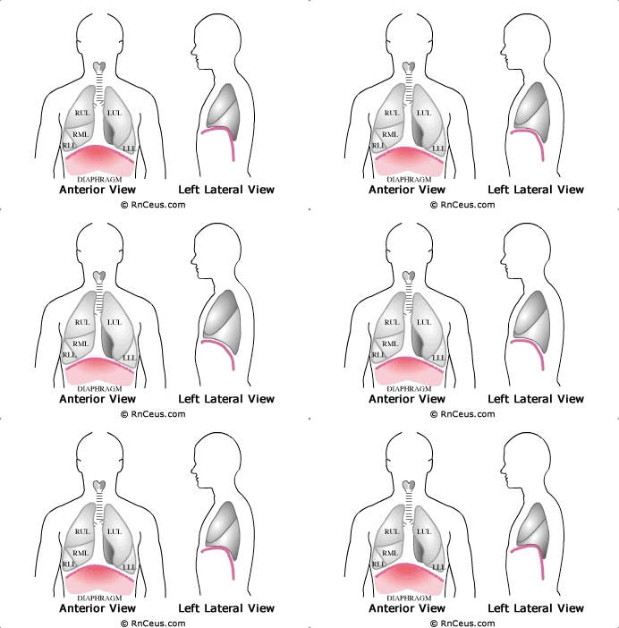
The illustration shows the movement of the diaphragm during the respiratory cycle.
- Watch as the diaphragm contracts, forcing the abdominal viscera downwards as the chest expands from top to bottom.
- The diaphragm is relaxed during exhalation, curving up towards the deflating lungs, as the elastic tissue passively recoils.
- With this front and side view (added to the rear view seen on the Respiratory Structure page), we have now located all of the lobes of the lung.
Instant
Feedback:
The
diaphragm is the major muscle of respiration and is innervated by the phrenic
nerve.
©RnCeus.com
 The diaphragm, a dome shaped
structure that separates the thoracic and abdominal cavities, is the major muscle
of respiration. The phrenic nerve innervates the diaphragm. The external and internal intercostal muscles are located between the ribs, increasing the anterior-posterior diameter of the thoracic cavity. Breathing may need to be assisted by other muscles, known as secondary or accessory muscles of respiration located in the neck and upper chest These muscles may include the parasternal, scalene, sternocleidomastoid, trapezius, and pectoralis muscles. Accessory respiratory muscles do not function during normal ventilation, but may be needed in some respiratory disorders.