Nonalcoholic Fatty Liver Disease (NAFLD)
Definitions
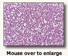
Nonalcoholic fatty liver disease (NAFLD) occurs in stages: simple fatty liver (NAFL), non-alcoholic steatohepatitis (NASH) and fibrosis.
- NAFL - nonalcoholic hepatic steatosis with no evidence of hepatocellular injury is mostly benign.
- NASH - nonalcoholic steatohepatitis is associated with a likely progression to cirrhosis, causing liver failure, the need for liver transplantation, and ultimately, hepatocellular carcinoma (HCC) (Rada 2020).
- Fibrosis - Scarring that compromises the circulation, lobar structure and hepatic function is known as cirrhosis.
Prevalence
- Children - NAFLD is the most common cause of chronic liver disease in children in the United States (Schwimmer 2006).
- Close to 10 percent of U.S. children ages 2 to 19 had NAFLD NAFLD has become more common in children in recent decades, in part because childhood obesity has become more common.
- About 23 percent of those with excess fat in the liver had NASH.
- NAFLD and NASH are more common in older children than in younger children.
- NAFLD is more common in boys than in girls. However, among children with
- NAFLD, girls and boys are equally likely to have NASH.
- Ethnic distribution of childhood NAFLD and NASH: Hispanic, Asian American, Caucasian and least common in African American children (Schwimmer 2006).
- Adult incidence
- NAFLD prevalence: 10-30% of U.S. population (17% in Framingham study)
- Nonalcoholic Steatohepatitis (NASH) accounts for one third of NAFLD cases
- Occurs in up to 3-5% of the U.S. population
- Affects up to 66% of age >50 years with obesity or diabetes mellitus
- Frequent cause of mild Liver Function Test abnormalities
- Most common cause of mildly abnormal ALT and AST in U.S. (accounts for up to 51% of cases)
- Most common cause of cryptogenic Cirrhosis (U.S. adult)
- By 2030, projected U.S. prevalence 100 Million cases, will become the top indication for liver transplant (Moses 2021).
 Etiology
- NAFLD - "insulin resistance leads to an increased influx of free fatty acids (FFA) into the liver. This happens due to the failure of insulin to suppress the hormone-sensitive lipase, causing more FFA to be released from the adipose tissue. Also, elevated insulin levels and insulin resistance promote continuous synthesis of triglycerides in the liver. These two sources of triglycerides result in accumulation of lipids in the hepatocytes causing macrovesicular hepatic steatosis" (Antunes (2021).
- NASH - "evolution from NAFLD caused by a second "hit." The evidence behind this second insult in not conclusive, but the most acceptable theories involve oxidative stress, specific cytokines, plus lipopolysaccharides.
 Free fatty acids and hyperinsulinemia potentiate lipid peroxidation and the release of hydroxy-free radicals, directly injuring the hepatocytes by recruiting neuroinflammatory mediators. Chronic liver injury over time will lead to activation of stellate cells, creating a potential for hepatic fibrosis" (Antunes (2021).
Signs and Symptoms (Moses 2021)
- Signs - Hepatomegaly in about 50%
- Symptoms
- Asymptomatic in most cases
- Fatigue
- Malaise
- Right upper quadrant pain
Diagnostics (Moses 2021)
- AST/ALT ratio
- NAFLD Fibrosis Score
- Magnetic Resonance Elastography Score
- Liver biopsy
Management (Moses 2021)
- Weight Reduction (consider bariatric surgery)
- Avoid hepatotoxins
- Maximize glucose control
- Lipid reduction prn
- Hypertension control
References
Antunes C. (2021). Fatty liver. StatPearls [Internet]. Retrieved November 13, 2021, from https://www.ncbi.nlm.nih.gov/books/NBK441992/.
Moses S. (2021). Nonalcoholic fatty liver. Family Practice Notebook. Retrieved November 13, 2021, from https://fpnotebook.com/gi/Lvr/NnlchlcFtyLvr.htm.
Rada P., González-Rodríguez, Á., García-Monzón, C., & Valverde, Á. M. (2020, September 25). Understanding lipotoxicity in NAFLD PATHOGENESIS: IS CD36 a key driver? Nature News. Retrieved November 13, 2021, from https://www.nature.com/articles/s41419-020-03003-w#citeas
Schwimmer JB, Deutsch R, Kahen T, Lavine JE, Stanley C, and Behling C. (2006). Prevalence of fatty liver in children and adolescents. Pediatrics. ;118(4):1388–1393
© RnCeus.com
 Etiology
 Free fatty acids and hyperinsulinemia potentiate lipid peroxidation and the release of hydroxy-free radicals, directly injuring the hepatocytes by recruiting neuroinflammatory mediators. Chronic liver injury over time will lead to activation of stellate cells, creating a potential for hepatic fibrosis" (Antunes (2021).