Central
Venous Pressure Monitoring
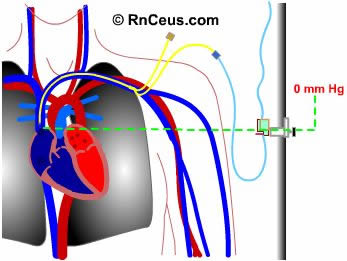
Central venous pressure is considered
a direct measurement of the blood pressure in the right atrium and vena cava.
It is acquired by threading a central venous catheter (subclavian double lumen
central line shown) into any of several large veins. It is threaded so that
the tip of the catheter rests in the lower third of the superior vena cava.
The pressure monitoring assembly is attached to the distal port of a multilumen
central vein catheter.
 |
Assisting with CVP placement
-
Adhere to
institutional Policy and Procedure.
-
Obtain history
and assess the patient.
-
Explain
the procedure to the patient, include:
- local anesthetic
- trendelenberg positioning
- draping
- limit movement
- need to maintain sterile
field.
- post procedure chest
X-ray
-
Obtain a
sterile, flushed and pressurized transducer assembly
-
Obtain the
catheter size, style and length ordered.
-
Obtain supplies:
-
Masks
-
Sterile
gloves
-
Line
insertion kit
- Heparin flush per policy
- Position patient supine
on bed capable of trendelenberg position
- Prepare for post procedure
chest X-ray
|
The CVP catheter is an important
tool used to assess right ventricular function and systemic fluid status.
- Normal CVP is 2-6 mm Hg.
- CVP is elevated by :
- overhydration which increases
venous return
- heart failure or PA stenosis
which limit venous outflow and lead to venous congestion
- positive pressure breathing,
straining,
- CVP decreases with:
- hypovolemic shock from hemorrhage,
fluid shift, dehydration
- negative pressure breathing
which occurs when the patient demonstrates retractions or mechanical negative
pressure which is sometimes used for high spinal cord injuries.
The CVP catheter is also an important
treatment tool which allows for:
- Rapid infusion
- Infusion of hypertonic solutions
and medications that could damage veins
- Serial venous blood assessment
Instant
Feedback:
The
CVP reading helps assess the function of the right ventricle and fluid status.
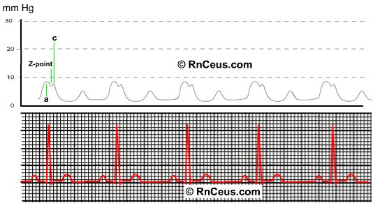
There are two ways to read a CVP
waveform:

1. Find the mean of the A wave.
- read the high point of the A
wave
- read the low point of the A
wave
- add the high point to the low
point
- divide the sum by 2
- the result is the mean CVP
The A wave starts just
after the P wave ends and represents the atrial contraction. The high point
of the A wave is the atrial pressure at maximum contraction. During the
A wave the atrial pressure is greater than the ventricular diastolic pressure.
At that point, the atrium is contracted, the tricuspid is open. Therefore,
the high point of the A wave closely parallels the right ventricular end
diastolic pressure. Remember, when the tricuspid valve is open and the right
ventricle is full, the ventricle, atrium and vena cavae are all connected.
Therefore, that point is the CVP.
2. Find the Z-point.
- Find the Z-point which occurs
mid to end QRS
- Read the Z-point
The Z-point coincides with the
middle to end of the QRS wave. It occurs just before closure of the tricuspid
valve. Therefore, it is a good indicator of right ventricular end diastolic
pressure. The Z-point is useful when A waves are not visible, as in atrial
fibrillation. (The c-wave occurs at closure of the tricuspid
valve. The crest of the c-wave is the atrial pressure increase caused by
the tricuspid valve bulging back into the atrium.)
Instant
Feedback:
Find
the Z-point to read a CVP waveform, when A waves are not visible.

