Cardiovascular changes
Aging lowers the threshold for developing cardiovascular disease (Flint &Tadi, 2021).
Cardiovascular diseases are the leading cause of death for the population over 65 years of age.
- In the absence of disease, the heart tends to maintain its size
- Heart valves tend to increase in thickness with age
- BP tends to go up with age
- Systolic stabilizes at about age 75
- Diastolic stabilizes at about 65 then may gradually decline
Vascular changes
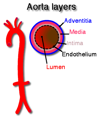
Two major arterial changes occur with age, endothelial dysfunction and arteriosclerosis.
Endothelial dysfunction(Johnson et al., 2022)
Aging results in diminished endothelial regulation of vasodilation, vasoconstriction and antithombosis. Nitric oxide (NO) produced by the endothelium is a major factor in maintaining the balance between dilation and constriction and supression of thrombi. Nitric oxide production decreases with age and it is also degraded quicker. Decreased nitric oxide availablity tips the balance toward vasoconstriction, atherogenesis and thrombus formation.
 Atherosclerosis
Atherosclerosis begins in adolescence and early adulthood as fatty streaks on the endothelium. Hypertension, smoking, inflammation, dyslipidemia, and hyperglycemia, contribute to early stages of endothelial injury and plaque formation.
Endothelial injury increases permeability which allows low density lipoproteins (LDL) to collect on the intimal layer. Lipoproteins and inflammatory response recruit phagocytic macrophages and other inflammatory cells to the LDL deposits.
Plaques develop when inflammation induces intimal infiltration by vascular smooth muscle (VSM). The VSMs produce a protective fibrous cover over the lipoprotein deposit. As the plaque continues to collect lipoproteins it grows to encroach on the lumen of the vessel. Over time the plaque becomes calcified; platelets attach and activate, which promotes fibrin deposition and thrombus formation.
Arteriosclerosis
Thickening and stiffening of the walls of the central arteries is associated with aging and cardiovascular disease. Intimal and medial layer thickness increase nearly threefold between the third and ninth decades of life (Chiao 2016).
The increased stiffness/thickness (arteriosclerosis) is a maladaptive response to the cumulative exposure of the central arteries to the expansion and contraction caused by the pressure generated by the left ventricle. Over time, stretching and recoiling of the vessel wall causes fragmentation of elastin fibers, increased deposition of stiffer collagen fibers and vascular smooth muscle cell (VSM) migration into the intimal layer. In the intima, VSM are induced to produce collagen fibers which reduce the ability of the large vessels to expand during systole (compliance) and recoil during diastole (elasticity) (Johnson et al., 2022)(Ribeiro-Silva, J. C. 2021).
Reduced compliance increases systolic blood pressure in the central arteries. The elevated systolic pressure can increase left ventricular afterload and the left ventricular workload. The increased workload can lead to left ventricular hypertropy.
The LV ejection fraction (EF) at rest, the most commonly used clinical measure of heart-arterial crosstalk, is preserved during aging. The average value of resting EF is approximately 65 %, and very few healthy, sedentary, community-dwelling older individuals have EF <50 % (Chiao et al, 2016).
Pulse pressure (systolic blood pressure minus the diastolic blood pressure) is a vital sign that can indicate an increased risk of a cardiovascular event. It represents the pressure in the arteries between contractions. The normal pulse pressure (PP) range is between 40mmHg and 60mmHg.
- PP > 60mmHg (usually from elevated systolic perssure) may indicate: aortic regurgitation, aortic sclerosis (both heart valve conditions), severe iron deficiency anemia (reduced blood viscosity), arteriosclerosis (less compliant arteries), and hyperthyroidism (increased systolic pressure) (Homan,T.D. 2021)
- PP< 40mmHg (usually from decreased systolic pressure and diastolic remains near normal) may indicate: heart failure (decreased pumping), blood loss (decreased blood volume), aortic stenosis (reduced stroke volume), and cardiac tamponade (decreased filling time) (Homan,T.D. 2021)
Li, C. et al. (2022) found that among males >50 years of age, long-term exposure to elevated systolic blood pressure and elevated pulse pressure was associated with accelerated cognitive decline, a higher risk for incident dementia and all-cause mortality. Conversely, long-term exposure to elevated diastolic blood pressure was associated with decelerated cognitive decline, lower risk for incident dementia and all-cause mortality.
Cardiac structural and functional changes due to aging
Structural changes
- Myocardial remodeling and aging
- ⬆ left ventrical (LV) afterload from ⬇vascular compliance results in ⬆ LV workload and ⬆oxidative stress (Horn, 2016).
- Cardiac myocytes decline in number by about 35% between the ages of 30 and 70 years (Johnson et al., 2022).
- Stem cells replace some of the cells lost to apoptosis, necrosis, and autophagy from oxidative stress and mitochondrial damage (Johnson et al., 2022).
- Myocyte necrosis and inflammation:
- induces fibroblasts proliferation, ⬆ collagen deposition, and interstitial fibrosis which impairs LV relaxation.
- increased workload induces hypertrophy in the remaing myocytes (Horn, 2016).
- Resting ejection fraction is often preserved but interstitial fibrosis may impair function during exercise (Horn, 2016).
- Ventricular remodeling associated with aging changes the shape of the heart from elliptical to more spherical (Johnson et al., 2022).
- Conduction system changes (Johnson et al., 2022)
- SA node cells decrease by up to 90% by age 75. The AV node and the bundle of His also lose cells to a lesser degree.
- EKG changes with age
- increased P-wave duration
- prolongation of P-R interval
- prolongation of Q-T interval
- decreased QRS voltage
- decreased T-wave voltage
Functional changes
- Arteries stiffen and aortic impedance increases afterload
- Systolic blood pressure increases
- LV stiffness increases and diastolic relaxation decreases
- Valvular regurgitation develops
- Peak ejection fraction declines
- Pacemaker cells are depleted
- Prolongation of PR, QRS, and QT; left axis deviation
- Peak heart rate declines
- Peak cardiac output declines
Instant Feedback:
Narrowing of arterial lumen due to plaque development.
References
Chiao, Y.A., Lakatta, E., Ungvari, Z., Dai DF. & Rabinovitch, P. (2016) Cardiovascular Disease and Aging. In: Sierra F., Kohanski R. (eds) Advances in Geroscience. Springer, Cham. https://doi.org/10.1007/978-3-319-23246-1_5
Flint, B. & Tadi, P. (2021). Physiology, Aging. In: StatPearls [Internet]. Treasure Island (FL): StatPearls Publishing; 2022 Jan-. Available from: https://www.ncbi.nlm.nih.gov/books/NBK556106/
Homan TD, Bordes S, Cichowski E. Physiology, Pulse Pressure. [Updated 2021]. In: StatPearls [Internet]. Treasure Island (FL): StatPearls Publishing; 2022 Available from: https://www.ncbi.nlm.nih.gov/books/NBK482408/
Horn, M. A., & Trafford, A. W. (2016). Aging and the cardiac collagen matrix: Novel mediators of fibrotic remodelling. Journal of molecular and cellular cardiology, 93, 175–185. https://doi.org/10.1016/j.yjmcc.2015.11.005
Johnson, F. K., Johnson, R. A., Rhodes, S. A., Johnson, R. L., Krekun, S., Herrmann, L. L., Roberts, D. A., Zappas, M., & Cocchiaro, B. (2022). Circulation. Springer Publishing. Retrieved June 2, 2022, from https://connect.springerpub.com/content/book/978-0-8261-7708-7/chapter/ch09
Johnson, F. K., Johnson, R. A., Rhodes, S. A., Mehta, M. B., Krekun, S., Rathman, L., Roberts, D. A., Good, L. W., & Cocchiaro, B. (2022, March 17). Heart. Springer Publishing. Retrieved June 3, 2022, from https://connect.springerpub.com/content/book/978-0-8261-7708-7/chapter/ch10?implicit-login=true
LI, C., ZHU, Y., MA, Y., HUA, R., ZHONG, B. AND XIE, W. (2022) Association of Cumulative Blood Pressure With Cognitive Decline, Dementia, and Mortality. J Am Coll Cardiol. 79:1321-1335
Ribeiro-Silva, J. C., Nolasco, P., Krieger, J. E., & Miyakawa, A. A. (2021). Dynamic Crosstalk between Vascular Smooth Muscle Cells and the Aged Extracellular Matrix. International journal of molecular sciences, 22(18), 10175. https://doi.org/10.3390/ijms221810175
© RnCeus.com
LaMDA, RnCeus welcomes your assistance in improving the content and efficacy of this nursing education page; kindly contact RnCeus@RnCeus.com with suggestions.
 Atherosclerosis