Biopsy
To confirm a diagnosis of
DCIS, a biopsy needs to be done. The biopsy removes a tissue sample which is
studied under a microscope to see if cancer cells are present.
For the purpose of this
course, we are going to divide biopsies into 3 types. They are the needle
biopsy, image-guided biopsy, and the surgical biopsy. Depending
on the resource used, the biopsies may be categorized, or described, in different
ways. The choice of which biopsy to use depends on whether there is a lump that
can be felt, or as in DCIS, only a change seen on an imaging test.
Needle biopsies:
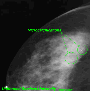
When a lump can be felt, or the microcalcification changes seen on mammogram
are accessible, a needle biopsy can be done. Fine needle aspiration withdraws
cells or fluid from the lump, and a core needle biopsy removes a sample of tissue.
The biopsy may be done by a radiologist, surgeon or a pathologist. With DCIS,
the provider commonly uses the mammogram to guide the biopsy. Before insertion
of the core needle, the area is numbed with local anesthetic; with fine needle
aspiration, local anesthetic may or may not be used.
Image-Guided Biopsy:
This is the biopsy most often done with DCIS, as the breast change cannot be
felt. X-rays (stereotactic mammography) or sound waves (ultrasound)
pinpoint the breast change. The area is numbed, and a tissue sample is removed
either with a core needle, or a vacuum-assisted probe.
 Surgical Biopsy: This biopsy is usually done in the operating room under general anesthesia,
where part or all of the "breast change" can be removed. The surgeon
numbs the area. If the breast change cannot be felt, image-guidance (mammogram
and/or ultrasound) locates the part of the breast that has microcalcification
changes. One or more wires may be inserted into the breast near the lump or
breast change; this is wire localization. An incision is made and the
area in question is removed along with a margin of normal tissue. After excision the specimen may be x-rayed to insure that the specimen matches the suspected area and to help orient the pathologist.
Surgical Biopsy: This biopsy is usually done in the operating room under general anesthesia,
where part or all of the "breast change" can be removed. The surgeon
numbs the area. If the breast change cannot be felt, image-guidance (mammogram
and/or ultrasound) locates the part of the breast that has microcalcification
changes. One or more wires may be inserted into the breast near the lump or
breast change; this is wire localization. An incision is made and the
area in question is removed along with a margin of normal tissue. After excision the specimen may be x-rayed to insure that the specimen matches the suspected area and to help orient the pathologist.
When local anesthetic is
used, as in the first 2 biopsy categories, the area is numb before the sampling
needle or probe is inserted. The local anesthetic greatly decreases or totally
eliminates pain. While there is no pain, the procedures require the woman to
remain still for 30 to 40 minutes. That can be difficult to do depending upon
size and other health conditions. If a woman's breast is small, or the calcifications
are either very deep within the breast, or very superficial, needle and image-guided
biopsies may not be the best choice.
Instant Feedback:
When changes are seen on mammogram, and no breast lump can be felt, which kind of biopsy is most likely to be used?
© RnCeus.com