Breast anatomy
Breast Anatomy
For more than 160 years, anatomical illustrations of the duct system of the female breast have been based upon the work of Sir Astley Pastor Cooper. Sir Cooper's skill, persistence, and natural deductive facility allowed him to describe the anatomy of the breast with a significant degree of fidelity.
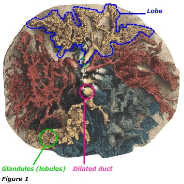
 Figure 1 depicts a wax injection cast prepared by Sir Cooper. Sir Cooper injected cadaver ducts at the nipple with yellow, red, green, blue, and black wax. The colors allow the spatial identification of lobes and lobules associated with each duct (Cooper, 1845).
Unfortunately, the tools and processes available to Sir Cooper, i.e., necropsy, boiling, and injection with hot wax, appear to have induced an artifact that has only recently been revealed. The dilated ducts located directly below the nipple have been presumed to be anatomically correct and have come to be known as lactiferous sinuses. Lactiferous sinuses were believed to be milk reservoirs important to effective suckling. Sir Cooper described the diameter of the milk reservoirs this way, "Their caliber is out of all proportion larger than that of the straight or mamillary tubes, and much larger than that of the milk tubes, which form their continuations" (Cooper, 1845).
Using high-resolution ultrasound technology, D. Ramsey et al. (2005) investigated the anatomy of the human lactating female breast. The investigators found:
- no evidence of lactiferous sinuses in the lactating breast
- myriad small milk ducts converging from the periphery to 4–18 main milk ducts at the base of the nipple
- the mean diameter of the ducts immediately below the nipple of a lactating breast ranged from 1.0-4.4 mm in diameter, similar to non-lactating breasts
- all ducts branched within the areolar radius, the first branch occurring 8.0 ± 5.5 mm from the nipple
- the diameter of the ducts increased at branching points
- the mean diameter of the ducts at the first branching distal to the nipple was 1.35 mm
- The ratio of glandular tissue to adipose is ≈ 2:1 in the lactating breast and ≈ 1:1 in the non-lactating breast
- excision of tissue within 30 mm of the nipple could significantly impact lactation
- ablation of only a few ducts can seriously impair lactation in some women
Breast anatomical structures
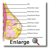
 The diagrams to the right show some basic anatomical structures of the breast. These structures are of interest in a discussion about DCIS.
Macro
• Typically, there is one breast on each side of the sternum, but breast tissue or nipples may occur anywhere along the embryonic milk lines.
• The adult female breast typically extends from the second rib superiorly to the 6th or 7th rib inferiorly and from the sternal border to the midaxillary line laterally. Rarely does breast tissue extend well beyond those landmarks.
• The breast develops within the superficial fascia and is retained in place by suspensory ligaments (Cooper's ligaments)
• The breast rests on the major chest muscle, the pectoralis major
• Fat surrounds and permeates the mammary lobes. Fat contributes to the size and shape of the breast.
Micro
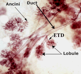
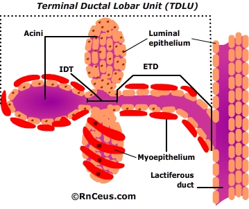
 Each breast, or mammary gland, contains 15-20 lobes, and each lobe comprises 20-40 terminal ductal lobular units (TDLU). The TDLU is the functional unit of the breast.
- TDLUs consist of:
- extralobular terminal duct (ETD), which attaches the lobule to the ductal system
- intralobular terminal duct (ITD) continues the duct system into the lobule
- clusters of 10-100 sac-like acini that open into the ITD.
- Acini and the terminal duct are the sources of milk production.
- "The epithelium throughout the ductal-lobular system is bilayered, consisting of an inner (luminal) epithelial cell layer and an outer ( basal ) myoepithelial cell layer."
- Visual, auditory, and areola stimulation triggers a neuroendocrine reflex which releases oxytocin from the posterior pituitary. Oxytocin travels in the blood to the mammary gland. It stimulates specific receptors on myoepithelial cells, causing them to contract and expel milk into the ducts and toward the nipple.
 Each lobe empties into a lactiferous duct.
- Lactiferous ducts merge into 5-10 main lactiferous ducts that open at the nipple.
- Most pathologic changes in the breast, including DCIS and invasive carcinomas, are believed to arise from the TDLU (Hall, 2020).
References
Cooper, A. (1845). The anatomy and diseases of the breast. Philadelphia: Lea & Blanchard.
Hall, J.E. & Hall, M.E. (2020). Guyton and Hall Textbook of Medical Physiology (Guyton Physiology) 14th Edition. New York, NY: Elsevier.
Ramsay, D.T., Kent, J.C., Hartmann, R.A. & Hartmann, P.E. (2005). Anatomy of the lactating human breast redefined with ultrasound imaging. J Anat. 206(6), 525-34.
 Figure 1 depicts a wax injection cast prepared by Sir Cooper. Sir Cooper injected cadaver ducts at the nipple with yellow, red, green, blue, and black wax. The colors allow the spatial identification of lobes and lobules associated with each duct (Cooper, 1845).
 Each breast, or mammary gland, contains 15-20 lobes, and each lobe comprises 20-40 terminal ductal lobular units (TDLU). The TDLU is the functional unit of the breast.
 Each lobe empties into a lactiferous duct.